Following directly on from a project to create an illustrated information sheet for radiotherapy patients (described on this webpage) Gill worked with scientists and clinicians at University College London (UCL) to create figures for a research study Patient Information Sheet (PIS). In this case, the patients taking part in the study would be very young children undergoing radiotherapy treatment for abdominal cancer and therefore, in the first instance, the PIS was designed for their parent or guardian. One of the scientists involved in this study had also been part of the illustrated information sheet project mentioned earlier and she was keen to use similar illustrations in the PIS.
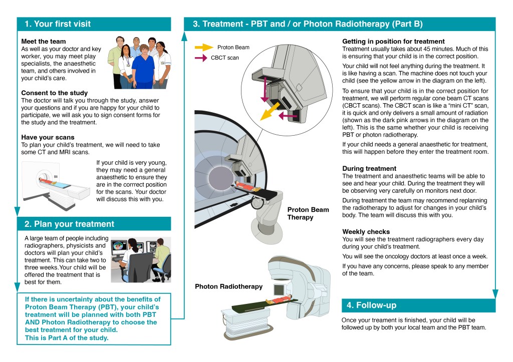
Gill met with the three scientists heading the study to discuss the figures that would be included in the PIS. The research study would involve patients receiving either Proton Beam Therapy (PBT) and/or Photon Radiotherapy during their treatment. It was decided that two A4-size figures would be required, one to show the treatment pathway for a patient (Figure 1) and the second to explain the difference between PBT and Photon Radiotherapy (Figure 2).
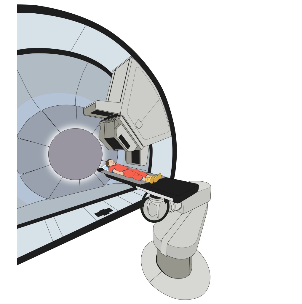
For Figure 1, Gill was able to revise and edit illustrations that she had created for existing projects to produce images of the radiotherapy team, the planning of the treatment and the MRI scanning. In the case of the latter image, it was just a case of replacing an adult patient with a child. The PBT machine, however, is very specialised, with only two in the country (one at UCL and the other at the Christie NHS trust in Manchester). In order to get a better idea of the different treatment machines and how they work, Gill visited the UCL radiotherapy centre to learn more about radiotherapy treatment. She was able to take photographs of both the PBT machine and the more conventional Photon Radiotherapy machine. She then used these photos as reference for the drawn illustrations that were used in the figure (as shown below).


Although Proton Beam Therapy is more accurate than Photon Radiotherapy, and so should cause less harm to healthy tissue surrounding any cancerous tumours, PBT is more sensitive to changes in the bowel contents and this can affect its accuracy. This is why scanning the patient just before treatment, to check for these changes, is so important. Figure 2, therefore, was designed to both explain the difference between PBT and Photon Radiotherapy, and to show how PBT is affected by changes in the bowel contents. The scientists were keen to use the same kind of torso X-section illustration that had been used in the illustrated information sheet for radiotherapy patients, and Gill used that image as the starting point for the range of illustrations shown below.


The illustrations were then combined with text to create the two figures, which involved a lot of communication between Gill and the UCL scientists and many iterative revisions of both text and image. The final versions of the two figures are shown below.

As mentioned earlier, these figures were designed for a PIS aimed at the parent or guardian of a young patient, rather than the patient themselves. Therefore, once the figures had been finalised, Gill revised the text to produce versions aimed at the patient themselves, that would be suitable for older children.
